PCR Testing
The Nuclein™ Hand-Held PCR Test is designed to be a disposable, all-in-one self-test for infectious disease diagnosis. It uses standard, real-time PCR, requires no technical expertise, and delivers battery-powered, sample-to-answer results in under one hour, without the need to mail in a sample. We are working tirelessly to make this available in 2021 to help with the COVID-19 crisis. Please note that this testing device will require FDA regulatory clearance and is meant to be used in consultation with a medical professional.
As shown on Our Solution page, we accomplish this by miniaturizing and integrating the same three steps that are used in traditional laboratories to perform PCR testing:
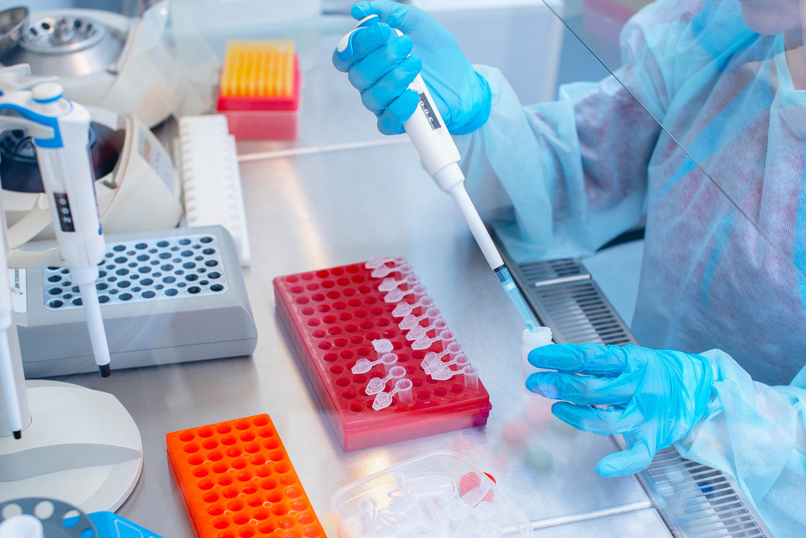
- Sample preparation
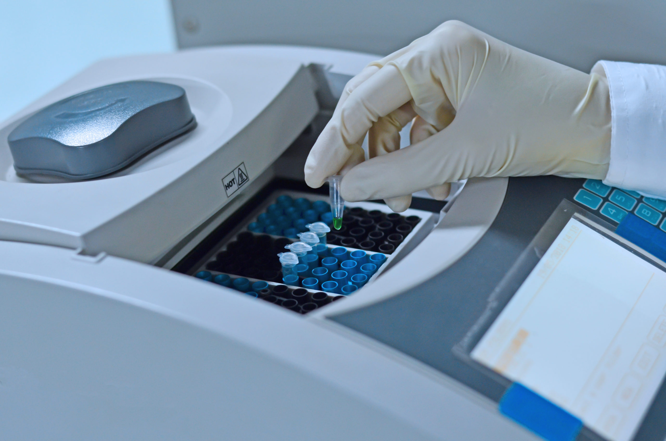
- Amplification
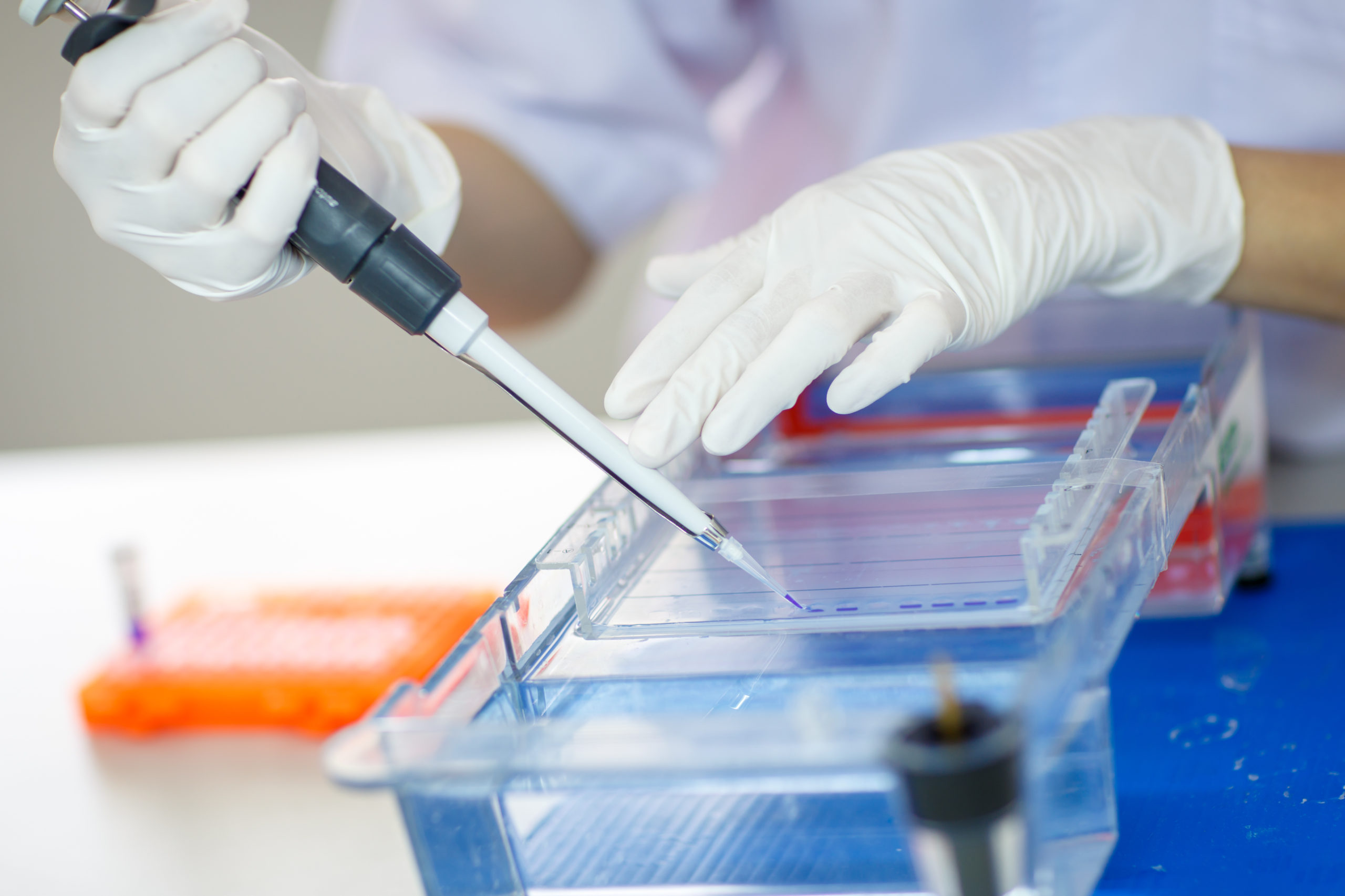
- Detection
A more technical explanation is presented below for those readers interested in additional details.
Sample Preparation
First, a raw biological sample from a patient such as saliva, blood, urine or other body fluids is processed to extract the nucleic acids. This sample preparation phase can be addressed in several ways, but principally, the sample is lysed, a process which kills and breaks open any cells, bacteria or viruses that are present.
Second, the natural enzymes present, which might otherwise degrade the nucleic acids in the lysate, are inactivated to produce a stable lysate and then depending on the application, some degree of purification may be performed to separate the nucleic acids from the other components of the lysate.
Finally, the purified nucleic acids may be concentrated in preparation for the next step, amplification.
Amplification
After lysis is complete, the targeted nucleic acids from the raw biological sample are ready to be amplified. When used for diagnostics, PCR provides information about whether a pathogen is present in a biological sample by amplifying only when the targeted nucleic acid sequence specific to that pathogen is present. The PCR reaction chemistry makes use of short strands of nucleic acids called primers, which are complementary to (or match) the pathogen’s sequence. When the primers bind to the nucleic acids in the reaction, an included enzyme called a polymerase can begin the process of amplification. It is important to note that the PCR reaction does not amplify (or make more of) the entire pathogen, only a small, non-infectious portion of its genetic sequence so that it can be identified.
DNA is a polymer made up of many small subunits called nucleotides. In a PCR reaction, the polymerase acts to assemble the nucleotides into new DNA strands that match the original. To achieve the exponential growth seen during the amplification phase, PCR uses multiple cycles of heating and cooling, called thermocycling. At high temperatures, heat forces each double-stranded DNA molecule apart into two single-stranded DNA molecules in a process called denaturation. As the two single strands begin to cool, the primers can anneal to them and the polymerase assembles the free nucleotides into new complementary strands. With each cycle, the new strands again separate and the amount of target DNA doubles. This repeated doubling allows a very small amount of nucleic acids to increase exponentially and makes detection possible.
Detection
For PCR diagnostics, one of the ways to detect the amplified nucleic acid is called real-time detection. Real-time PCR means that the amplification can be monitored as the reaction progresses (for example, every cycle), and a result can be reported as soon as the amplification is detected.
To obtain higher specificity for diagnostics, sequence-specific probes called hydrolysis probes can be used so that a signal is only detected if the probe and the amplified nucleic acid are complementary. Hydrolysis probes work by combining a fluorophore and a quencher. The quencher normally blocks the fluorescent signal from the fluorophore, but once the probe binds to the amplified nucleic acid sequence it matches, the fluorophore and quencher become separated, revealing the fluorescence of the fluorophore.
In this way, if the specific target of interest (e.g., nucleic acids from a bacteria or virus) is present, the fluorescent signal from the reaction increases each cycle of amplification. A device sensor that measures the amount of fluorescence in the reaction can report the presence of the bacteria or virus of interest as soon as the fluorescence signal becomes large enough to be detected.


